
Cranial nerves are 12 pairs of nerves originating from the brain, controlling sensory, motor, and parasympathetic functions. They connect the brain to muscles and sensory organs, enabling essential bodily functions like vision, hearing, and swallowing. Understanding their structure and function is crucial for accurate neurological assessment and diagnosis.
1.1 Overview of Cranial Nerves
Cranial nerves are 12 pairs of nerves originating from the brainstem, controlling vital sensory, motor, and parasympathetic functions. They regulate vision, hearing, smell, taste, and motor activities like chewing, swallowing, and facial expressions. Each nerve has specific roles, such as the olfactory nerve for smell and the optic nerve for vision. These nerves connect the brain to sensory organs, muscles, and glands, enabling communication and coordination. Their complex pathways and functions make them essential for diagnosing neurological disorders, as damage can lead to significant deficits. Understanding their anatomy and functions is fundamental for accurate clinical assessment and patient care.
1.2 Importance in Neurological Assessment
Cranial nerve assessment is critical in neurological evaluation, as it provides insights into the brain’s control over sensory and motor functions. These nerves connect the brain to sensory organs and muscles, enabling essential functions like vision, hearing, and swallowing. Damage to cranial nerves can indicate underlying neurological conditions, such as stroke or tumors. Accurate assessment helps identify deficits early, guiding diagnosis and treatment. It also aids in monitoring disease progression and recovery. A thorough understanding of cranial nerve function is vital for clinicians to detect abnormalities and ensure comprehensive patient care. This makes cranial nerve evaluation a cornerstone of neurological practice.

Cranial Nerve Assessment Preparation
Preparation involves positioning the patient comfortably, ensuring a quiet environment, and gathering essential tools like a Snellen chart, olfactory test materials, and a flashlight for pupil assessment.
2.1 Patient Positioning and Environment
Patient positioning is critical for accurate cranial nerve assessment. The patient should sit comfortably in a chair, 1 meter away from the examiner, at the same eye level. Ensure the environment is quiet and well-lit to minimize distractions. For certain tests, like hearing assessments, a soundproof room may be necessary. Positioning varies slightly depending on the nerve being tested: for example, the patient should be seated upright for olfactory nerve testing and asked to close their eyes during smell identification. Proper positioning ensures accurate results and patient cooperation during the examination process.
2.2 Necessary Equipment and Tools
The cranial nerve assessment requires specific tools to evaluate sensory and motor functions accurately. Essential equipment includes a Snellen chart or hand-held acuity card for vision testing, a tuning fork for hearing assessments, and non-pungent odor bottles for olfactory nerve evaluation. A penlight is used for pupillary reflex testing, while a cotton swab or soft brush assesses facial sensation. Gloves and protective equipment ensure hygiene. Additional tools like a tongue depressor and neurological hammer may be needed for specific reflex tests. Having all necessary tools prepared ensures a smooth and efficient assessment process, allowing the examiner to thoroughly evaluate each cranial nerve function.
Sensory Functions Assessment
This section evaluates the sensory functions of cranial nerves I, II, and VIII, using methods like odor identification, vision charts, and hearing tests to detect deficits.
3.1 Cranial Nerve I (Olfactory Nerve)
The olfactory nerve (CN I) is responsible for transmitting sensory information related to smell. Assessment involves presenting the patient with non-pungent odors, such as vanilla or coffee, and asking them to identify the scent. Each nostril is tested separately, with the patient’s eyes closed to ensure reliance on olfaction. This evaluation helps detect deficits in smell perception, which may indicate neurological conditions or trauma affecting the olfactory pathways. Accurate documentation of the patient’s ability to identify odors is crucial for diagnosing impairments in this cranial nerve function.
3.2 Cranial Nerve II (Optic Nerve)
The optic nerve (CN II) transmits visual information from the retina to the brain. Assessment involves evaluating visual acuity, visual fields, and pupillary reactions. Using a Snellen chart or handheld acuity card, each eye is tested separately to measure sharpness of vision. Confrontation testing checks peripheral vision, while the swinging flashlight test assesses pupillary light reflex, helping identify defects like afferent pupillary defects. These tests are essential for diagnosing conditions such as optic neuritis or glaucoma. Accurate documentation of findings aids in localizing lesions and guiding further neurological evaluation.

Motor Functions Assessment
Motor functions are evaluated by assessing cranial nerves controlling eye movements, facial expressions, and swallowing. Tests include tracking eye movements, facial muscle strength, and tongue protrusion to identify deficits.

4.1 Cranial Nerves III, IV, and VI (Eye Movements)
Cranial nerves III (oculomotor), IV (trochlear), and VI (abducens) control eye movements. Assessment involves evaluating the patient’s ability to track an object in six cardinal directions using eye movements. The examiner moves an object horizontally, vertically, and diagonally, observing for smooth, coordinated movements. Nystagmus or inability to move eyes in specific directions may indicate nerve dysfunction. Pupil reactions, such as the pupillary light reflex, are also tested for CN III. Abnormalities in these nerves can cause diplopia, strabismus, or limited gaze. Accurate testing ensures early detection of neurological deficits affecting vision and ocular function.

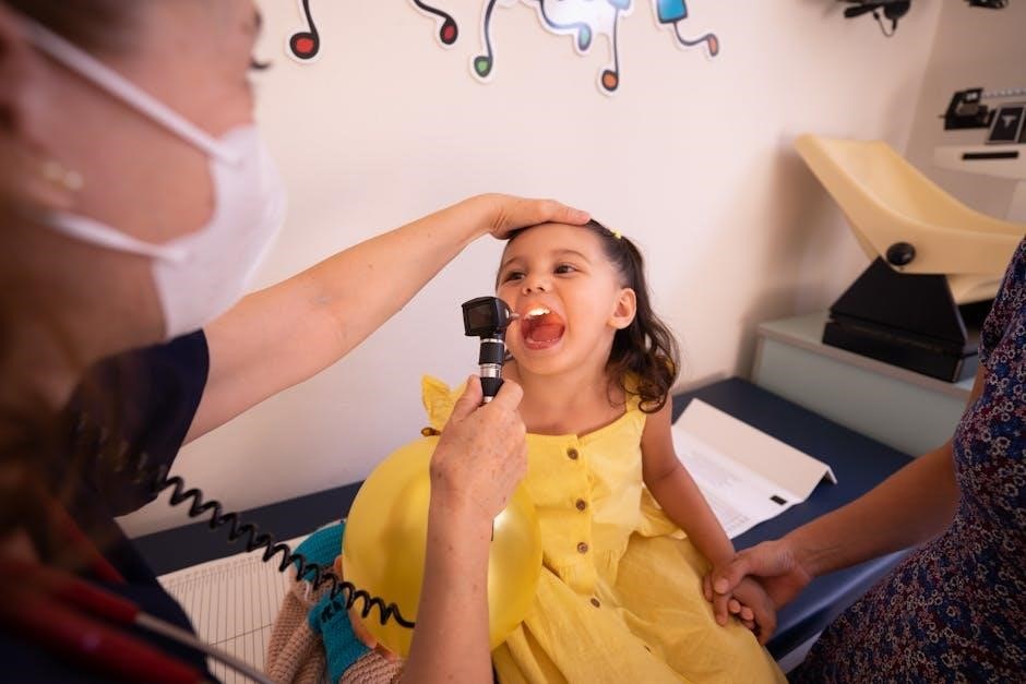
4.2 Cranial Nerve V (Trigeminal Nerve)
Cranial Nerve V, the trigeminal nerve, is responsible for facial sensation and motor functions. It has three branches: ophthalmic, maxillary, and mandibular. Sensory assessment involves lightly touching the face with a soft object to check for sensation in all branches. The corneal reflex is tested by gently touching the cornea with a cotton swab, eliciting a blink. Motor function is evaluated by asking the patient to clench their jaw and protrude it against resistance, testing the masseter and temporalis muscles. Abnormalities may indicate nerve damage, leading to sensory loss or weakened chewing ability.
4.3 Cranial Nerve VII (Facial Nerve)
Cranial Nerve VII, the facial nerve, manages facial expressions and taste. Motor function is assessed by having the patient smile, frown, and raise eyebrows, noting symmetry and strength. Each side should be tested separately to identify unilateral weakness. For taste, place sweet, sour, salty, or bitter substances on the tongue and ask for identification. Any asymmetry, weakness, or taste dysfunction may signal nerve damage or conditions like Bell’s palsy. This evaluation is crucial for accurate neurological assessments and diagnoses.
4.4 Cranial Nerve VIII (Vestibulocochlear Nerve)
Cranial Nerve VIII, the vestibulocochlear nerve, is responsible for hearing and balance. Assessment includes gross hearing tests like the Rinne and Weber tests. The vestibular-ocular reflex is evaluated by observing eye movements during head turning. Patients may be asked to march in place with eyes closed to test balance. Abnormal findings, such as nystagmus or hearing loss, can indicate nerve damage or conditions like Ménière’s disease. Accurate evaluation of this nerve is essential for diagnosing auditory and vestibular disorders, ensuring proper neurological assessment and management.
4.5 Cranial Nerves IX and X (Glossopharyngeal and Vagus Nerves)
Cranial Nerves IX and X are crucial for swallowing, taste, and vocal cord function. Assessment involves examining the gag reflex, uvular elevation, and the patient’s ability to swallow. The gag reflex is tested by stimulating the pharynx. Patients are asked to say “ahh” to inspect soft palate movement. Taste sensation on the posterior tongue is evaluated for CN IX. CN X regulates vocal cord function, tested through speech and cough reflexes. Abnormalities may indicate dysphagia or neurological conditions, making this assessment vital for diagnosing swallowing and respiratory disorders.
4.6 Cranial Nerve XI (Accessory Nerve)
Cranial Nerve XI, the accessory nerve, controls the sternocleidomastoid and trapezius muscles, essential for neck rotation, shoulder elevation, and head movement. Assessment involves testing muscle strength and movement. Patients are asked to shrug shoulders and turn their head against resistance. Weakness or paralysis in these muscles may indicate nerve damage, often due to trauma or neurological conditions. Accurate evaluation of CN XI is vital for diagnosing motor deficits and guiding rehabilitation or surgical interventions to restore functional mobility in the neck and shoulder region.
4.7 Cranial Nerve XII (Hypoglossal Nerve)
Cranial Nerve XII, the hypoglossal nerve, governs tongue movements, including protrusion, lateral deviation, and manipulation of food during chewing and swallowing. Assessment involves observing tongue protrusion and movement. Weakness or paralysis may cause tongue deviation toward the affected side, indicating a lesion. Testing includes asking the patient to stick out their tongue and move it side to side. Atrophy or fasciculations may also be noted. Accurate evaluation of CN XII is essential for diagnosing swallowing and speech disorders, ensuring proper neurological and rehabilitative care for patients with suspected hypoglossal nerve dysfunction.

Clinical Application and Documentation
Accurate documentation of cranial nerve assessment findings aids in identifying patterns, tracking progress, and informing treatment plans. Standardized methods ensure consistency and reliability in clinical records, enhancing patient outcomes.
5.1 Interpreting Assessment Results
Interpreting cranial nerve assessment results involves comparing findings to normal function to detect deficits. Abnormalities like diminished reflexes or impaired sensory responses may indicate specific nerve lesions or systemic issues. For example, weak facial muscles suggest a potential CN VII lesion, while speech difficulties could point to CN IX or X involvement. Accurate interpretation requires a thorough understanding of each nerve’s functions and their corresponding clinical signs. Proper documentation and correlation with other neurological findings ensure precise diagnosis and appropriate intervention. Regular practice and clinical experience refine interpretation skills, enhancing patient care outcomes;
5.2 Documentation Best Practices
Accurate and detailed documentation is crucial for cranial nerve assessments. Use standardized templates to record findings systematically, ensuring clarity and completeness. Document both positive and negative results, noting any abnormalities or deficits. Avoid vague terms; instead, use specific descriptors like “impaired” or “absent.” Include baseline measurements for future comparisons. Store records securely, adhering to confidentiality guidelines. Regularly review and update documentation to reflect patient progress or changes. Clear documentation facilitates effective communication among healthcare providers, ensuring continuity of care and informed decision-making. It also serves as a legal record of the assessment process and findings.